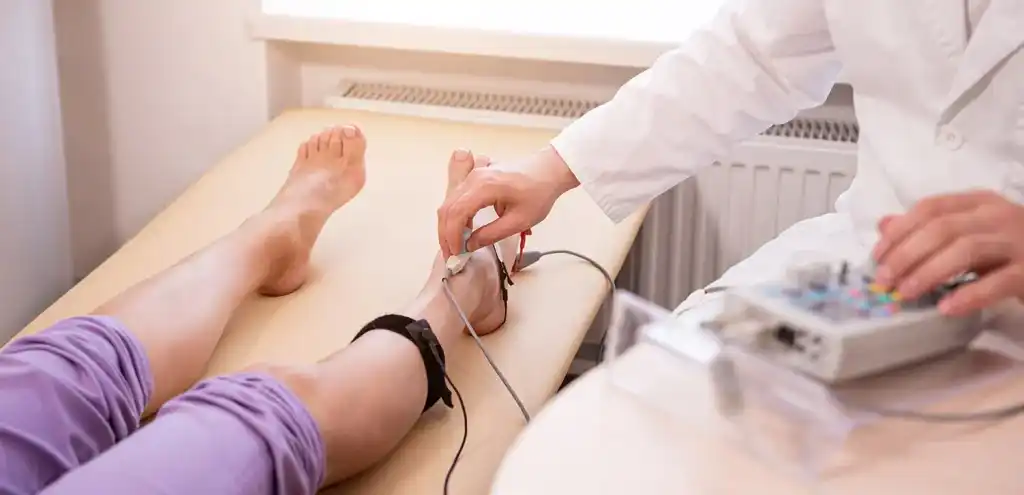
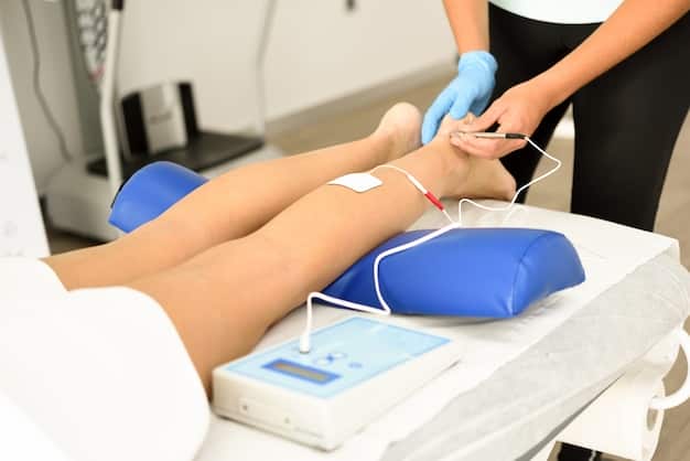
Nerve Conduction Studies (NCS/ENG)
Electromyography (EMG)
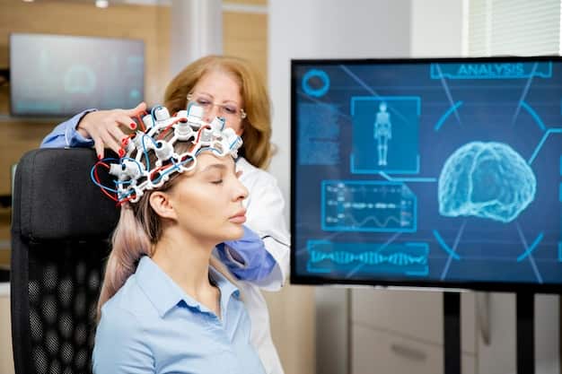
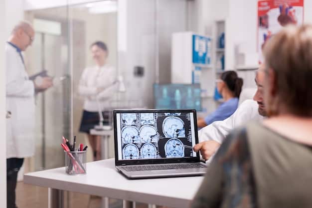
Electroencephalography (EEG)
Evoked Potentials (EP)
Visual Evoked Potentials (VEP)

Somatosensory Evoked Potentials (SEP)

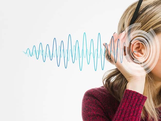
Auditory Evoked Potentials (AEP)
Memory Testing

Botulinum Toxin Injections
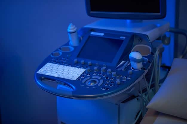
Ultrasound of the Neck Vessels